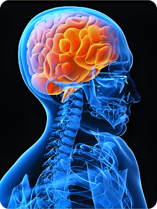
 A cerebral or brain aneurysm is where the vessel in the brain develops a “balloon-like” dilation as a result of a weakening within the inner layer of a blood vessel wall. This dilation can become extremely thin and rupture without warning as it fills with blood. This “balloon-like” dilation can place pressure on a nerve or surrounding brain tissue. If it leaks or ruptures, this can lead to blood spilling into surrounding tissue, known as a hemorrhage.
Cerebral aneurysms are primarily found in the base of the skull. Most brain aneurysms are congenital, meaning, an inborn abnormality in the structure of the artery. There is a higher incidence of aneurysms in people with connective tissue disorders and polycystic kidney disease, and certain circulatory disorders, such as arteriovenous malformations.
Diagnosis:
Many cerebral aneurysms often go unnoticed until they rupture or are detected by brain imaging like a computed tomography (CT) angiogram scan. There are several gold standard diagnostic tests that are available to help provide more information about the aneurysm. These tests also help determine the best course of treatment.
The majority of these tests are usually obtained after a hemorrhage in order to confirm the diagnosis of a cerebral aneurysm as well as identify the shape and size of the aneurysm. For example,
- Intracerebral angiogram: a dye test to help analyze arteries and identify the degree of narrowing or obstruction of the artery or blood vessel in the brain
- Magnetic resonance imaging (MRI): medical imaging technique that uses a magnetic field and computer generated radio waves to create detailed images of the brain
- Magnetic resonance angiography (MRA): medical imaging that produces more detailed images of blood vessels
Cerebrospinal fluid analysis can also be ordered if a ruptured aneurysm is suspected. This is a minimally invasive procedure which requires a local anesthetic to be used and a small amount of fluid is obtained by a surgical needle. This test will detect any bleeding or brain hemorrhage.
Treatment:
Every aneurysm is different and therefore treatment plans are individualized. Considerations for treatment take into account the size, type, and location of the aneurysm. Patient’s age, medical history and risk and benefits for treatment are also considered.
Not all cerebral aneurysms rupture and some small aneurysms may be continuously monitored for growth or acute onset of symptoms.
There are two surgical options available:
Microvascular clipping: This procedure primarily involves directly cutting off the blood flow to the aneurysm. Under anesthesia, a section of the skull is removed and the aneurysm is located. The neurosurgeon uses a microscope to find vessels that feed the aneurysm and a small, metal, clothespin-like clip is placed at the base of the aneurysm, stopping all blood flow to that aneurysm.
Endovascular embolization: This is an alternative to surgery where the neurosurgeon inserts a hollow plastic tube into an artery and threads it until the brain aneurysm is reached. Detachable coils (small spirals of platinum wire) or small latex balloons are passed through the catheter to go inside the aneurysm. The coils and or small latex balloons subsequently fill the aneurysm and block it from filling with blood. The aneurysm is thus destroyed.