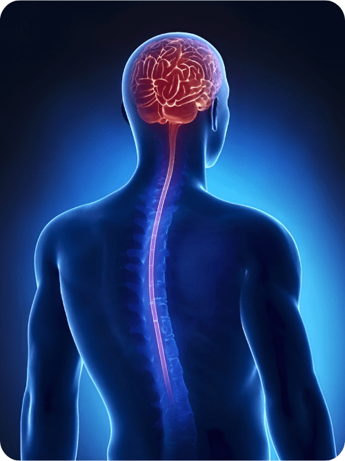
 Spinal stenosis occurs when there is a narrowing in the spaces within your spine that applies pressure on your spinal cord and nerves. This typically occurs in the neck and lower back area and are usually related to age-related wear and tear.
Cervical stenosis: narrowing of the spine in the neck area
Symptoms: numbness/tingling/weakness in hand, arm, foot, or leg; neck pain, loss of bowel or bladder function (severe cases)
Lumbar stenosis: narrowing of the spine in the lower back (most common form of spinal stenosis)
Symptoms: numbness/tingling/weakness in foot or leg; pain or cramping in one or both legs; back pain
Causes can include:
- Normal wear and tear associated with age.
- Osteoarthritis can cause the development of bone spurs that can grow into the canal.
- Herniated disks: The soft cushions between your spinal column (vertebrae) start to dry out with age. Cracks within the disks can escape and press on the spinal cord and nerves.
- Thickened ligaments: The strong cords that hold the bones of your spine together can also stiffen with time and age. These thickened cords can then press onto the spinal cord.
- Tumors: abnormal growth of cells in the spinal cord
Spinal injuries: Any trauma that can cause dislocations or fractures to the vertebrae. This fractures can damage the spinal cord.
Diagnosis
Dr. Tsimpas will ask about your symptoms, your past medical history, and conduct a physical examination. There are several diagnostic imaging studies that may be obtained to help identify the root cause of your symptoms.
- X-ray: An X-ray of your back can reveal bony changes, such as bone spurs that may be narrowing the space within the spinal canal.
- Magnetic resonance imaging (MRI): medical imaging technique that uses a magnetic field and computer generated radio waves to create detailed images of the spine and possibly identify any structural abnormalities that could be pressing onto the spinal cord
- Computerized Tomography (CT) or CT myelogram: This non-invasive imaging can help obtain different angles of the spine to outline the spinal cord, nerves. Herniated disks and bone spurs can also be identified. The CT myelogram uses dye (contrast) to help visualize structures of the spine.
Treatment
Treatment for spinal stenosis highly depends on the location of the stenosis and severity of your presenting symptoms.
If symptoms are mild, your doctor may monitor your condition and possibly recommend rehabilitation therapy.
Surgery is considered when other treatments have not improved your symptoms or function.
Laminectomy: removing part of the affected vertebra and will relieve the pressure on the nerves
Laminotomy: a small incision is made into the bony area of the spine in order relieve the pressure on the nerves
Laminoplasty (cervical stenosis only): space within the spinal canal is made by creating a hinge on the bony area of the spine; metal hardware is placed to support the gap in the opened section of the spine.
Minimally invasive surgery: this type of surgery utilizes smaller incisions in order to cause less harm to nearby tissues
Discectomy (most common surgery for lumbar stenosis): portion or all of the disk is removed to alleviate the pressure on the affected nerve root
Spinal fusion: surgical technique where two or more joints are fused together to simulate the normal healing process of broken bones. This a permanent connection between two or more vertebrae of your spine and therefore eliminating motion between them.